AI System for Kidney Tumor Classification
October 2025
An advanced AI diagnostic support system designed to minimize unnecessary surgeries by accurately distinguishing malignant from benign renal tumors.
Overview
The primary goal of this project is to accurately distinguish between malignant (cancerous) and benign (non-cancerous) kidney tumors in CT scans. By achieving high diagnostic certainty, we aim to significantly reduce the rate of unnecessary surgical interventions and biopsies for patients with benign masses.
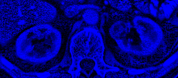
While Computed Tomography (CT) is the standard for diagnosis, creating AI that can efficiently process its massive 3D data is difficult. To solve this, we developed a system that acts like a "Synthetic Multi-Spectral Sensor." It breaks down complex medical images into three simplified visual channels-one for structure, one for density, and one for texture.
By analyzing these features separately, our solution achieves 97.0% precision while requiring a fraction of the computational power of traditional 3D systems.
The Challenge
Because standard CT scans obscure critical tissue textures, surgeons face diagnostic uncertainty, leading to a high rate of unnecessary, redundant surgical interventions for indeterminate kidney masses (with up to 30% ultimately found to be benign).
Our Solution
An Expert-Informed Multi-Spectral AI that breaks down monochromatic CT scans into three distinct bands (Structure, Density, Texture), exposing hidden malignancy signs to eliminate diagnostic ambiguity.
The Challenge: Diagnostic Ambiguity in Standard Imaging
Standard Computed Tomography (CT) imaging compresses vast amounts of radiodensity data into simplified grayscale imagery. This limitation obscures the subtle differences between benign and malignant renal masses, making visual diagnosis difficult for medical staff.
- Diagnostic Ambiguity: Traditional viewing windows often clip critical data extremes, erasing definitive visual signs of specific tumor types like Angiomyolipomas or Renal Cell Carcinomas.
- Hidden Micro-Textures: Crucial indicators of malignancy frequently reside in subtle, localized textures that standard viewing methods render invisible to the human eye.
- The Urologist's Dilemma: Because standard scans obscure critical tissue textures, surgeons are often forced to remove indeterminate kidney masses "just to be safe." This diagnostic uncertainty directly drives overtreatment: clinical data indicates that up to 30% of surgically removed small renal masses are ultimately found to be benign. From a hospital's perspective, this cautious approach results in high liability, wasted Operating Room (OR) time, and avoidable patient harm.
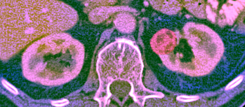
The Solution: Multi-Spectral Image Transformation
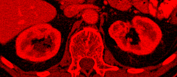
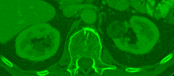
To overcome these clinical limitations, our platform transforms a standard monochromatic CT scan into a rich, multi-spectral representation. By separating the image into three distinct analytical channels, we expose critical data previously hidden from view:
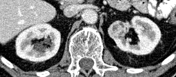
Acts as the structural foundation, establishing the general shape and physiological context of the organ.
Captures extreme tissue densities outside the standard clinical view, crucial for identifying fatty tissues and calcifications.
Applies non-linear filters to isolate micro-textures, separating cohesive benign tissue from chaotic, malignant growths.
By mapping these biological features into three color channels, we triple the amount of useful information the AI receives without slowing it down.
Targeted Neural Attention
We engineered a highly efficient method to direct the AI's diagnostic focus. By systematically shifting the color properties of suspected tumor regions, we naturally draw the network's attention to pathological areas without adding computational overhead.
The diagnostic model is trained in two distinct phases:
- Phase 1: Guided Discovery - The system learns to anchor essential tumor characteristics utilizing structural masks, rapidly accelerating baseline accuracy.
- Phase 2: Independent Adaptation - The model adapts to locate and classify tumors autonomously, relying entirely on learned synthetic textures and densities within raw, unsegmented data.
Results and Phased Implementation
Our implementation strategy is structured in two sequential phases to guarantee clinical validity and operational robustness:
- Phase 1 (Current): Base Model Training - The system was initially trained and validated on a large, open-source database containing roughly 600 annotated CT scans, successfully proving the efficacy of the multi-spectral architecture.
- Phase 2 (Planned): Clinical Fine-Tuning - The architecture will be fine-tuned against a proprietary hospital database of approximately 2,000 diverse CT scans. This phase adapts the system to real-world, institution-specific imaging variances prior to clinical deployment.
Based on the initial Phase 1 data, the classifier achieved state-of-the-art predictive performance while remaining highly efficient.
Operational Benefits
- Fast and Accessible: Because it processes data efficiently, the system can run on standard hospital computers without requiring expensive, specialized hardware.
- Highly Reliable: Important features defining malignancy are never lost during the data preparation phase.
- Explainable AI: The platform offers visual verification. Doctors can manually review the synthesized Red, Green, and Blue channels to understand and confirm why the AI made its decision.
By translating complex medical reasoning into the data preparation phase, we avoid the "Black Box" problem of AI. Our system provides transparent, accurate, and resource-efficient diagnostic support.
This solution serves as a robust tool designed to streamline surgical assessments, safely reduce unnecessary operations, and directly reinforce physician confidence.
Interested in a Custom AI Solution for Your Medical Challenges?
We specialize in developing AI systems for the medical field that combine technological innovation with a deep understanding of clinical needs. Let's create a solution together that will improve diagnosis, treatment, and quality of life for patients.
Contact Us Today